In a groundbreaking development for agricultural science and soil research, scientists have successfully integrated computed tomography (CT) scanning with advanced laser 3D reconstruction techniques to visualize root systems and soil structures with unprecedented clarity. This innovative approach promises to revolutionize our understanding of below-ground ecosystems, offering new insights into plant-soil interactions that could transform farming practices and environmental conservation efforts.
The technology combines high-resolution X-ray CT scanning – traditionally used in medical diagnostics – with laser-based 3D modeling to create detailed, three-dimensional representations of root architectures within their native soil environments. What sets this method apart is its ability to capture both the biological components (roots) and the physical soil matrix simultaneously without destructive sampling, preserving the intricate relationships between plants and their growing medium.
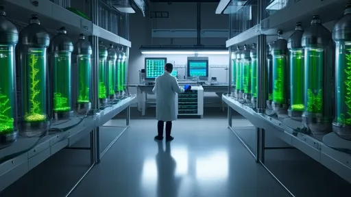
Researchers at several leading agriscience institutions have been developing this technique over the past five years, overcoming significant technical challenges related to differentiating root material from soil particles in CT images. The solution emerged through the application of specialized image processing algorithms that can distinguish organic matter from mineral components based on their density and structural signatures.
The implications for agricultural research are profound. For the first time, scientists can observe how root systems develop and interact with soil structures under various conditions – drought, different fertilizer applications, or varying soil compositions – all in real time and without disturbing the experimental system. This non-invasive approach allows for longitudinal studies that track changes over entire growing seasons.
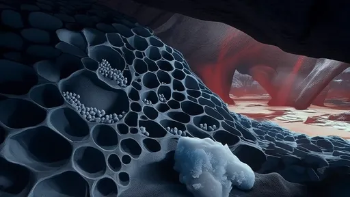
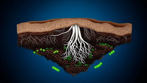
One particularly exciting application involves studying the rhizosphere – the narrow region of soil directly influenced by root secretions and associated microorganisms. The 3D reconstructions reveal how roots modify their immediate environment, creating complex networks of pores and aggregates that affect water retention, nutrient cycling, and microbial habitats. These findings could lead to more targeted soil management practices that optimize these natural processes.
The laser 3D reconstruction component adds another dimension to the technology. After CT scanning captures the internal structures, laser scanning creates precise surface models of soil samples or field plots. When combined, these datasets produce comprehensive digital twins of soil-root systems that can be rotated, sectioned, and analyzed from any angle. Researchers can virtually "dissect" these models to examine features that would be impossible to observe through physical dissection.
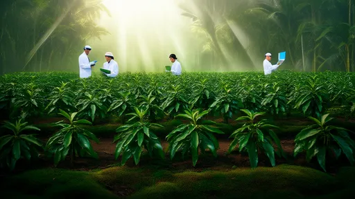
Field applications of this technology are already yielding valuable data. In one ongoing study of cereal crops, the imaging system has revealed previously unknown patterns of root growth in compacted soils. The 3D models clearly show how certain root varieties can navigate dense soil layers by finding and exploiting microscopic cracks and pores – information that could inform breeding programs for more resilient crop varieties.
The technology's precision extends to quantifying root characteristics that were previously difficult to measure accurately. Parameters like root diameter distribution, branching angles, and surface area can now be determined with sub-millimeter accuracy across entire root systems. This level of detail is helping researchers develop more accurate models of water and nutrient uptake by plants.
Beyond agriculture, the method shows promise for ecological restoration projects. Scientists studying native plant communities can now document how different species' root systems interact below ground, informing more effective planting strategies for habitat restoration. The ability to visualize soil structure changes over time also provides valuable data for assessing the effectiveness of various soil remediation techniques.
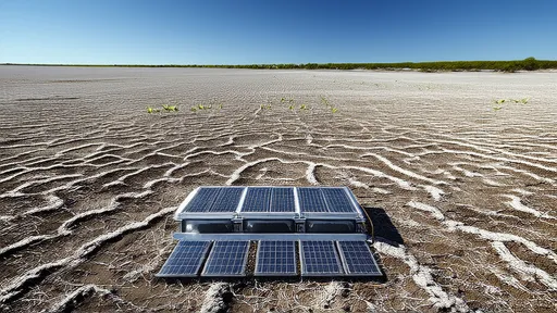
While currently used primarily in research settings, the developers envision broader applications for this technology. Future iterations could include portable field units that would allow farmers to assess soil structure and root health directly in their fields. There's also potential for integrating the technology with precision agriculture systems, providing data to optimize irrigation and fertilization at the root zone level.
The team is currently working to improve the speed and resolution of the scanning process while reducing costs. Early versions required several hours to scan a single soil core, but recent advancements have cut this time significantly. The goal is to make the technology accessible to a wider range of research institutions and eventually to agricultural extension services.
As climate change alters precipitation patterns and soil conditions worldwide, understanding below-ground plant adaptations becomes increasingly crucial. This novel imaging approach provides the tools to study these changes at unprecedented scales and resolutions. The detailed 3D models serve not only as research tools but also as powerful educational resources, making the hidden world beneath our feet visible and comprehensible.
The integration of medical imaging technology with agricultural science exemplifies the innovative cross-pollination of disciplines driving modern research. What began as a diagnostic tool for human health is now shedding new light on plant health and soil ecosystems, demonstrating how technological advancements in one field can yield unexpected benefits in another.
Looking ahead, researchers anticipate that the data generated by this method will feed into larger digital agriculture initiatives, contributing to more sustainable and productive farming systems. As the technology matures and becomes more widely adopted, it may well become a standard tool for soil scientists and agronomists, fundamentally changing how we study and manage the critical interface between plants and soil.

By /Aug 14, 2025

By /Aug 14, 2025

By /Aug 14, 2025
By /Aug 14, 2025

By /Aug 14, 2025

By /Aug 14, 2025

By /Aug 14, 2025
By /Aug 14, 2025

By /Aug 14, 2025
By /Aug 14, 2025
By /Aug 14, 2025

By /Aug 14, 2025

By /Aug 14, 2025

By /Aug 14, 2025

By /Aug 14, 2025
By /Aug 14, 2025

By /Aug 14, 2025
By /Aug 14, 2025
By /Aug 14, 2025
By /Aug 14, 2025